Only HD Wallpapers
⌕
Log in
Log in
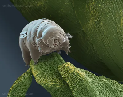
← Tardigrade on Leaf
Related to Tardigrade on Leaf
Page 3 of 5
plant
25.6k
microscopic
381
science
2.4k
leaf
9.1k
biology
615
tardigrade
8
Microscopic Cells — Blue
1920 × 1200
leaf vein structure close-up
1600 × 1200
bacterial cells under microscope
1440 × 900
Leaf Surface under Microscope
2848 × 2136
Microscopic Diatoms
1600 × 1200
Dividing Cells — Blue, Scientific
1024 × 768
Dividing Cells
1280 × 1024
Leaf Vein Structure
1600 × 1000
Close-up of Leaf Veins — Green, Natural, Photograph, Texture, Yellow
1600 × 963
microscopic view of plant cells
2088 × 1550
Pollen Grain
1600 × 1200
The Nitrogen Cycle Diagram
1309 × 801
Leaf shape and venation guide
2163 × 1977
Virus Infecting Cells
1920 × 1200
3D cell division illustration
1280 × 800
Cell division in progress
1600 × 1200
Jellyfish eggs under microscope
1600 × 1200
close-up of insect mouthparts
909 × 1500
Red Blood Cells — Clinical, Blood Plasma
3360 × 1050
green leaves with chemical structures
1440 × 900
Virus-infected cell under microscope
3000 × 2395
Green Plant Leaves — Calm, Photograph, Drops
2560 × 1600
Virus Particles under Microscope — Blue, Clinical
1680 × 1050
Bacteria under Microscope — Black
1400 × 1050
← Previous
Page 3 of 5
Next →